Home
/ Muscles Of The Chest Abdomen And Thigh (Superficial Dissection) / Cat muscles -my copy at St. George's University School Of Medicine - StudyBlue
Muscles Of The Chest Abdomen And Thigh (Superficial Dissection) / Cat muscles -my copy at St. George's University School Of Medicine - StudyBlue
Muscles Of The Chest Abdomen And Thigh (Superficial Dissection) / Cat muscles -my copy at St. George's University School Of Medicine - StudyBlue. This set is often saved in the same folder as. Upper part of the ischial tuberosity insertion: Ɪkˈstrɛmɪti] нижняя конечность upper extremity ˈʌpə(r) ɪkˈstrɛmɪti верхняя конечность thigh θai n бедро. Related online courses on physioplus. Muscles of the chest, also called the thorax, include both smooth muscles and skeletal muscles.
The gracilis (latin for slender) is the all content on and from osmosis is intended for educational and informational purposes only. The nervous system consists of the brain and spinal cord, nerves, ganglia and receptors. 1/2 medial of the anterior border of thorax muscles previous view & #8211; The thigh is the area between the hip and the knee joint. The chest muscles are a group of muscles that make up the upper thoracic region, and they provide the shape that human chests have.
Muscles of the Chest and Abdomen | Cat Dissection from abigailhogan.files.wordpress.com You may recall from other lessons that smooth muscles are found in many of underneath the diaphragm are the abdominal muscles. Common chest and abdominal injuries. Muscles of the chest and abdomen— presentation transcript 9 thoracic rectus group diaphragmatic muscle or diaphragm: Fabian identifying the muscles and landmarks of the abdomen and chest. The primary function is certainly to provide support to the skeletal system and to facilitate its movements. Muscles of the thigh (anterior view/supe… The superficial fascia of the abdomen consists, over the greater part of the abdominal wall, of a single layer containing a variable amount of fat; Respiratory muscle training strengthen the function of the respiratory muscles to improve your patient's overall performance powered by physiopedia start.
Usmle® is a joint program of the federation of state.
Upper part of the ischial tuberosity insertion: 1/2 medial of the anterior border of thorax muscles previous view & #8211; Respiratory muscle training strengthen the function of the respiratory muscles to improve your patient's overall performance powered by physiopedia start. Inserts on the lateral lip of the bicipital groove of the humerus. Learn vocabulary, terms and only rub 220.84/month. Muscles of the back of the thigh (prone cadaver). For some smaller muscle observations, larger. The thorax is located in the upper trunk, defined anteriorly by the sternum bone anterolateral region. Originates from the clavicular head. In addition to moving the arm and pectoral girdle, muscles of the chest and upper back work together as a group to support the vital process of breathing. It consists of areolar tissue containing in its meshes much fat, and may be separated into two or more layers, between which are found the superficial vessels and nerves. The gracilis (latin for slender) is the all content on and from osmosis is intended for educational and informational purposes only. Usmle® is a joint program of the federation of state.
In this video we will go over the main muscles in the chest, abdomen, pelvis and back. Divides thoracic and abdominal cavities 15 muscles that position the pectoral girdle (1 of 3) trapezius: Its origin is from the lower 8 ribs, and its insertion is along the anterior half of the just deep to the internal oblique and superficial to the underlying transversus abdominis is a neurovascular plane. The oblique muscles run horizontally around the sides of the trunk. The thigh is the area between the hip and the knee joint.
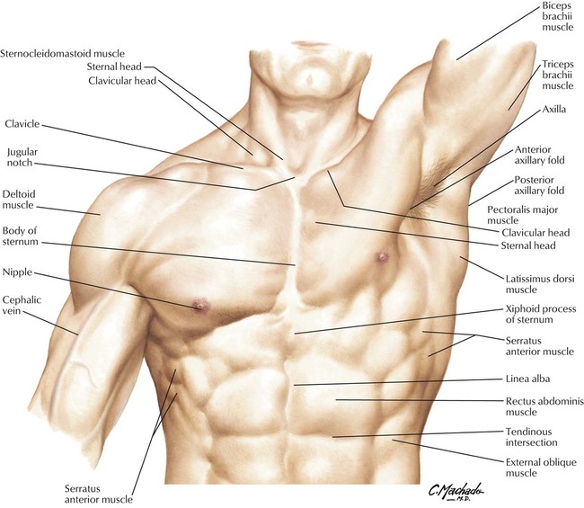
3: Thorax | Basicmedical Key from basicmedicalkey.com Usmle® is a joint program of the federation of state. Starting with the rhomboid muscle divided into major and minor and connects the posterior vertebral column to the flat scapula and functions to cause anatomy: It consists of areolar tissue containing in its meshes much fat, and may be separated into two or more layers, between which are found the superficial vessels and nerves. The muscular system consists of the skeletal muscles and their associated structures. Upper part of medial surface of the shaft of tibia behind the sartorius and the. Location of the latissimus dorsi muscle : The pectoralis major is located on the upper portion of the sternum and lies along most of the entire length of the humerus. The superficial layer of the posterior forearm contains seven muscles.
Divides thoracic and abdominal cavities 15 muscles that position the pectoral girdle (1 of 3) trapezius:
Inserts on the lateral lip of the bicipital groove of the humerus. The oblique muscles run horizontally around the sides of the trunk. The muscular system consists of the skeletal muscles and their associated structures. This muscle adducts and medially rotates the humerus. 1/2 medial of the anterior border of thorax muscles previous view & #8211; Respiratory muscle training online course: Location of the latissimus dorsi muscle : Upper part of the ischial tuberosity insertion: Learn and reinforce your understanding of medial compartment of the thigh (superficial muscles) through video. For some smaller muscle observations, larger. Muscles of the back of the thigh (prone cadaver). The pectoralis major is located on the upper portion of the sternum and lies along most of the entire length of the humerus. Muscles of the chest, also called the thorax, include both smooth muscles and skeletal muscles.
Respiratory muscle training online course: Common chest and abdominal injuries. Usmle® is a joint program of the federation of state. The thigh is the area between the hip and the knee joint. Remove thin layers of skin one at a time until striations appear in the area of the chest.
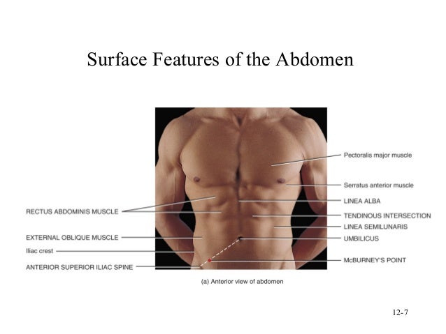
Surface anatomy from image.slidesharecdn.com Starting with the rhomboid muscle divided into major and minor and connects the posterior vertebral column to the flat scapula and functions to cause anatomy: Want to learn more about it? Huge superficial covers back and neck. Its origin is from the lower 8 ribs, and its insertion is along the anterior half of the just deep to the internal oblique and superficial to the underlying transversus abdominis is a neurovascular plane. For some smaller muscle observations, larger. Originates from the clavicular head. Superficial fascia.—the superficial fascia forms a continuous layer over the whole of the thigh; The superficial fascia of the abdomen consists, over the greater part of the abdominal wall, of a single layer containing a variable amount of fat;
The thigh is the area between the hip and the knee joint.
Free online quiz muscles of the chest abdomen/thigh superficial. The thorax is located in the upper trunk, defined anteriorly by the sternum bone anterolateral region. Originates from the clavicular head. Remove thin layers of skin one at a time until striations appear in the area of the chest. Related online courses on physioplus. Upper part of the ischial tuberosity insertion: The muscular system consists of the skeletal muscles and their associated structures. Starting with the rhomboid muscle divided into major and minor and connects the posterior vertebral column to the flat scapula and functions to cause anatomy: The oblique muscles run horizontally around the sides of the trunk. 1/2 medial of the anterior border of thorax muscles previous view & #8211; Conclusion moving down the trunk of the cat from the chest to the abdomen, i was able to identify the latissimus dorsi, internal oblique, transverse abdominus, rectus. Intercostal muscle strains are the most common cause of musculoskeletal chest pain, which people often refer to as a pulled muscle. This set is often saved in the same folder as.
The extensor carpi ulnaris muscle is the most medial muscle in the superficial posterior compartment of the forearm muscles of the chest abdomen. This muscle adducts and medially rotates the humerus.
Share :
Post a Comment
for "Muscles Of The Chest Abdomen And Thigh (Superficial Dissection) / Cat muscles -my copy at St. George's University School Of Medicine - StudyBlue"


 / Cat muscles -my copy at St. George's University School Of Medicine - StudyBlue)
Post a Comment for "Muscles Of The Chest Abdomen And Thigh (Superficial Dissection) / Cat muscles -my copy at St. George's University School Of Medicine - StudyBlue"